

The transmission electron microscope is the best choice in diagnosing diseases
The chemical diagnostic methods are less effective for surgical pathologists, and the repercussions are that they become interested in transmitting electron microscopy (TEM) for the purpose of getting additional diagnostic insights. Even though there are different types of electron microscopes (analytic, scanning, and high-voltage), the diagnosis in this field depends primarily on the transmission electron microscope.
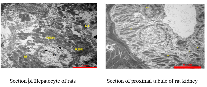
Consequently, the best alternative that the pathologists need to consider is a transmission electron microscope due to its high-resolution ability to view extracellular and intracellular structures. Microscopy techniques, like using a transmission electron microscope, reveal high-resolution images of minute intracellular and extracellular structures suchjson as collagen fibers, mitochondria, endoplasmic reticulum, and actin filaments.
Biopsies and surgical tissue are suitable for electron microscopic examination, covering many tissue and cell types. The best results are obtained from fresh extirpated specimens rather than formaldehyde-fixed or paraffin-embedded tissues. Fixation was carried out by immersion of the specimens in fixative glutaraldehyde solution and then post-fixing the specimens in osmium tetroxide, dehydration in graded alcohols, and finally implanting in epoxy resin. Staining of thick epoxy sections, measuring 1 µm, is accomplished by utilising a toluidine blue solution. The sections were then contrasted with uranyl acetate and lead citrate in sequence and cut into thin sections of 90 to 120 nm by means of a diamond knife and viewed under the electron microscope..
Ultrastructural investigations are a must to diagnose a number of neoplastic and non-neoplastic diseases, in particular, renal, glomerulopathies, peripheral neuropathies, and striated muscle (skeletal and heart).
Referenses
1- Erlandson RA. Raven Press; New York: 1994. Diagnostic Transmission Electron Microscopy, with Clinicopathological, Immunohistichemical, and Cytogenetic Correlations.
2-Ladanyi M, Lui MY, Antonescu CR. The der(17)t(X;17) (p11;q25) of human alveolar soft part sarcoma fuses the TFE3 transcription factor gene to ASPL, a novel gene at 17q25. Oncogene. 2001;20:45–57.